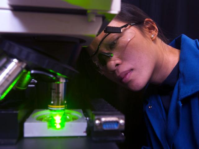
Leading the way to a better tomorrow
For over 20 years, our program has been equipping doctoral students to make a difference – take a look at how we create a critical bridge between academia and the biotech industry.
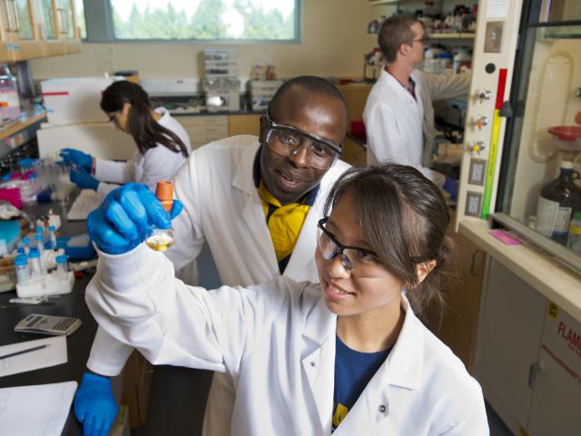
We offer opportunities for both students and corporate employees to pursue a doctoral degree with an emphasis in biotechnology. Learn about these opportunities and how to enroll.
There are several ways you can partner and support our program to help advance the future of biotechnology – from internships to donations, support keeps our program cutting-edge.